
DMEK Procedure
DMEK Surgery
Have you been told that you have endothelial dysfunction?
Patients with corneal endothelial dysfunction used to have few options for long-term, good visual acuity. As few as 10-15 years ago, people who presented with these diseases could only undergo full-thickness penetrating keratoplasty (PKP). Today, however, patients with trauma to the endothelial layer or Fuch’s dystrophy can undergo procedures that only remove the damaged endothelium.
In a short amount of time, deep lamellar endothelial keratoplasty (DLEK), Descemet-stripping endothelial keratoplasty (DSEK) and Descemet’s membrane endothelial keratoplasty (DMEK) have become the options of choice for partial corneal transplant surgeries. DMEK is the newest procedure, which is known as Descemets Membrane Endothelial Keratoplasty (DMEK), and is the latest innovation in minimally invasive corneal transplantation. It involves transplanting a delicate sheet of corneal cells.
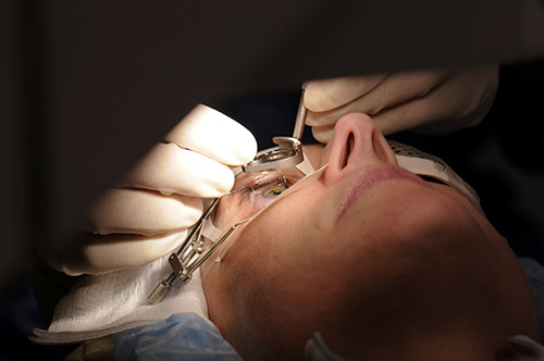
What happens in DMEK cornea surgery?
DMEK or Descemet’s membrane endothelial keratoplasty is the newest surgical technique for treatment of the endothelial corneal disease. The inner layer of the patient’s own cornea is stripped and a very thin membrane of a donor cornea is inserted. An air bubble is inserted into the eye to cause the thin membrane to uncurl and to position it behind the patient’s own cornea. The advantage of the DMEK technique is there are a potentially higher percentage of patients attaining best-corrected vision of 20/20 after this type of surgery.
The rejection rate for the DMEK may be reduced compared to a full thickness transplant. In DMEK cornea surgery, the patient’s original cornea is left mostly intact, so it’s not immediately apparent that he’s had a transplant. It may also be possible for the patient to attain 100% vision within a few weeks to a few months of surgery.
Data courtesy of Cornea Research Foundation
What is the endothelium and how does it work?
Since we mention endothelial dysfunction we should elaborate a bit more what it is!
The cornea is the clear dome that makes up the front part of your eye. It is about 1/20th of an inch thick and is composed of five layers: the epithelium, Bowman’s layer, the stroma, Descemet’s membrane and the endothelium. The epithelium is a thin surface layer. The stroma is composed mostly of proteins called collagen. This layer needs to stay clear for good vision.
The endothelium is a single layer of cells coating the inside portion of the cornea. Its job is to provide nutrients to the cells in the stroma and to make sure that the stroma has just the right amount of water. Too much water in the stroma can cause swelling of the cornea and decreased vision. When the endothelium fails then the stroma will have too much water and will not be clear. The endothelium cannot regenerate.
Question: Why would we select DMEK for a patient with endothelial dysfunction?
Answer: DMEK leaves the patient’s cornea closer to its original condition than any other transplant technique.
Benefits of DMEK
- 2.8 mm or smaller corneal incision
- No increase in corneal thickness
- No refractive shift from donor graft
- Ability to pre-operatively plan astigmatism management to reduce or eliminate pre-operative cylinder
- No changes or guesses for “fudge factors” with IOL calculations